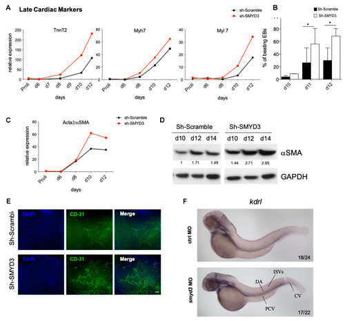
SMYD3 modulates late cardiovascular markers. (A) Cardiac markers, TnnT2, Myh7 and Myl7, were measured by qRT-PCR at d0, d6, d8, d10 and d12 of mESCs differentiation. Data were normalized to GAPDH. One representative experiment is shown; n = 2. (B) Beating EBs were counted at d10, d11, d12 of EBs formation in Sh-Scramble and Sh-SMYD3. Significance was calculated by 2-way Anova, followed by Bonferroni post hoc test. * p < 0.05 value for the significance is shown in the plot. n = 3. (C) αSMA mRNA levels were measured by qRT-PCR as in (A). One representative experiment is shown: n = 2. (D) Western blot analysis of αSMA protein extracts of Sh-Scramble and Sh-SMYD3 EBs at d10, d12, d14. GAPDH serves as a loading control. Normalized band intensity in immunoblots is reported below signals. (E) Immunofluorescence microscopy was performed on d10 EBs with antibodies against the endothelial marker CD31. DAPI stains nuclei. Scale bar: 100 μm. (F) kdrl expression was analyzed at 48 hpf, in standard control and smyd3 morphants. Lines show posterior cardinal vein, dorsal aorta, inter-segmental vessels and caudal vein (PCV, DA, ISVs, CV, respectively).
|
