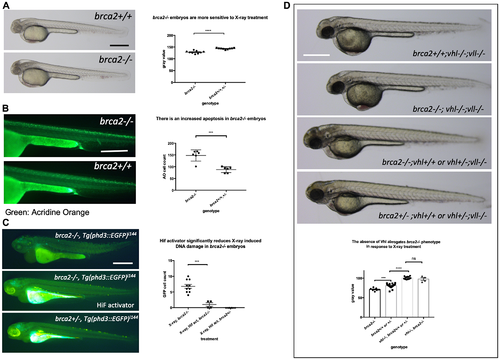
The elevated Hif level in the vhl–/– mutants alleviates cell deaths induced by the mutation in brca2. (A) brca2–/– embryos were more sensitive to X-ray treatment in comparison to their siblings demonstrating the increased cell death in CNS. ****p < 0.0001, unpaired t-test. (B) The Acridine Orange staining confirmed the increased apoptosis in the brca2–/– embryos. ***p < 0.001, unpaired t-test. (C) X-ray treatment induced a few EGFP positive cells in the brca2–/– embryos, indicating the loss of both vhl alleles in these embryos. On the other hand the EGFP positive cells were hardly observed in their siblings. The Hif activator treatment suppressed the appearance of EGFP positive vhl mutant cells in the brca2–/– embryos. ***p < 0.001, one way AVOVA. (D) brca2–/– embryos were more sensitive to X-ray treatment in comparison to their siblings. However, when the vhl –/–;vll –/– mutations are introduced in the brca2–/– embryos, increased apoptosis in the CNS of brca2–/– embryos was suppressed and the brca2–/– embryos were indistinguishable from their siblings. ***p < 0.001, ****p < 0.0001, nsp > 0.05, one way ANOVA. Scale bars: 0.5 mm.
|
