|
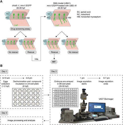
Automated small-molecule compound screening follows a two-step workflow. (A) Schematic representation of the axonal phenotype in chodl−/−; mnx1:GFP embryos (left) with stalled axons at the horizontal myoseptum (HM), and zebrafish model of SMA (UBA1 model, right) showing abnormal motor axons, as used in drug screening. Chemical compounds that rescue the chodl−/−; mnx1:EGFP axonal phenotype are tested again using the UBA1 model. SC, spinal cord; NC, notochord. (B) Schematic representation of timeline and workflow of the experimental protocol. At 8 hpf, zebrafish eggs are arrayed in a 24-well plate (six eggs per well) and incubated in drug solution overnight. The next day (day 2) the 28-30 hpf embryos are moved from the 24-well plate into a 96-well plate (three embryos per well) followed by automated imaging using the VAST BioImager. In a single imaging session, between 16 and 20 chemical compounds can be tested with six embryos per compound.
|