FIGURE

Figure 2

ID
ZDB-FIG-210518-34
Publication
Wu et al., 2021 - Automatic wavelet-based 3D nuclei segmentation and analysis for multicellular embryo quantification
Other Figures
All Figure Page
Back to All Figure Page
Figure 2

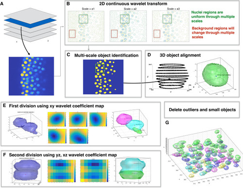
Overview of wavelet-based nuclei segmentation method. (A) Import 3D raw image files. (B) After importing 3D images, 2D continuous wavelet transform with appropriate wavelet scale factors are applying to each 2D z-stack. (C) 2D segmentation masks are obtained by applying multi-scale object identification to wavelet coefficient maps from previous step. (D) In 3D object alignment step, connected identified 2D masks from neighboring slices forms a 3D foreground object. (E) First division step if more than one center or center shift on neighboring z slice wavelet coefficient maps. (F) Second division step in z direction if multiple centers found on yz or xz wavelet coefficient maps. (G) Delete 3D nuclei voxels smaller then nuclei size × 0.2.

Expression Data

Expression Detail
Antibody Labeling
Phenotype Data

Phenotype Detail
Acknowledgments
This image is the copyrighted work of the attributed author or publisher, and ZFIN has permission only to display this image to its users. Additional permissions should be obtained from the applicable author or publisher of the image. Full text @ Sci. Rep.