
Fig. 5
- ID
- ZDB-FIG-210513-5
- Publication
- Banavar et al., 2021 - Mechanical control of tissue shape and morphogenetic flows during vertebrate body axis elongation
- Other Figures
- All Figure Page
- Back to All Figure Page
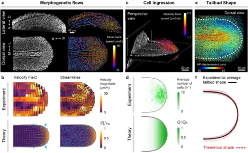
Morphogenetic flows and tissue shape during zebrafish posterior body axis elongation. (a), Confocal lateral and dorsal sections of nuclei (gray) in posterior tissues of a zebrafish embryo at the 10 somite stage (left). Results of nuclear (cell) tracking showing a subset of identified nuclei (blue) and associated tracks (trajectories), color-coded according to their mean speed. (b), Experimentally measured and theoretically obtained velocity field and streamlines of the morphogenetic flows in ventral tissues. (c), Perspective 3D view of a confocal stack through elongating posterior tissues, showing a D–V section at the boundary between dorsal and ventral tissues and a AP section far from the posterior end. Tracks of cells entering ventral tissues from DM tissues are shown and color-coded according to their maximal speed. (d), Measured frequency map of cells entering the MPZ from DM tissues and theoretically assumed spatial distribution of the same quantity, namely Q(x, y). (e), Dorsal view of cell (nuclear) trajectories in posterior tissues, color-coded according to the magnitude of their AP displacement. The boundary of ventral tissues is shown as a thick dashed line and determines the shape of the elongating posterior body. (f), Comparison of experimentally measured and theoretically predicted shapes of the posterior elongating tissue. The simulation results in (b,d,f) are all for the same parameters (𝜆𝜇/𝜆𝑄=1.5, 𝜎A/𝑃C=0.1, 𝜎P/𝑃C=0.02, 𝜎C/𝑃C=0.0001). |