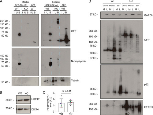
Procollagen processing controls. (A) Immunoblot of a GFP trap of media and lysate fractions of WT and giantin KO RPE1 cell cultures. Cells were either expressing GFP-COL1A1 or GFP alone as indicated. Blots show the input (I), unbound (U), and bound (B) fractions of the IP immunoblotted for GFP, pro-α1(I) N-propeptide (LF39 antibody), and tubulin (housekeeping). (B) Immunoblot of HSP47 and DIC74 (housekeeping) in WT and giantin KO RPE1 cell lysates. (C) Densitometry of the semiquantitative enhanced chemiluminescent immunoblots represented in A. HSP47 levels are normalized to DIC74. Each dot represents an independent biological replicate, and replicates are color coded between cell lines. Bars show median and interquartile range (n = 5 biological replicates). P value calculated with a Mann-Whitney U test. (D) Immunoblots of media (M) and lysate (L) fractions of WT and giantin KO RPE1 cell cultures stably expressing GFP-COL1A1 following treatment with DMSO (vehicle control), MG132, or bafilomycin (Baf). Blots are probed for GAPDH (housekeeping), GFP (GFP-COL1A1), p62 (positive control for bafilomycin), and pro-α1(I) (COL1A1) as indicated. A.U, arbitrary units.
|
