|
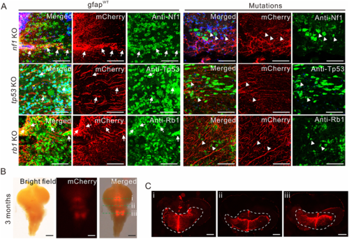
Disruption of tissue-specific genes in larvae and adult zebrafish. (A) Representative images of various single-deletion mutations, and gfapWT control in 3-month-old adult fish. The fluorescent radial glia phenotype was observed based on mCherry-labelled Gfap expression, which failed to co-localize with Nf1, Rb1 or Tp53 expression in brain tissues of transgenic zebrafish (arrowheads; right), whereas partial co-localizations of mCherry-labelled Gfap with Nf1, Rb1 or Tp53 expression were observed in gfapWT fish (arrows; left). Scale bars = 50 μm. (B) Merged bright and fluorescent images showing mCherry-labelled Gfap expression in the cerebellum and medulla in 3-month-old gfapWT fish. Scale bars = 500 μm. (C) Coronal sections of the images shown in i–iii. Robust mCherry-labelled Gfap expression was observed along the midline and dorsal lining of the fourth ventricle. Scale bars = 50 μm.
|