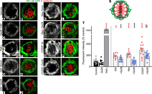
Qx28 protects hair cells by NF-κB pathway inhibition in vivo. Five dpf Tg(NFKB:EGFP) were treated with E3 water (A and B), Qx28 1 nM (C and D), TNF-α 20 ng/mL 30 minutes (positive control) (E and F), gentamicin (GM) 100 μM 30 minutes without (G and H) or with (I and J) Qx28, neomycin (Neo) 200 μM 5 minutes without (K and L) or with (M and N) Qx28, and CDDP 400 μM 2 hours without (O and P) or with (Q and R) Qx28. Animals were transferred to E3 water for 2 hours and immunostained for GFP (green) and otoferlin (red). The green fluorescence intensity was quantified using ImageJ and expressed as percentage from control (T). (S) Cartoon depicting a neuromast (top view): hair cells are in red, supporting cells are in gray, and mantle cells are in green. Results are expressed as mean ± SEM. Statistical analysis: 2-tailed Student’s t test, **P < 0.01, ***P < 0.001, ****P < 0.0001 versus ototoxin alone (black asterisks). One-way ANOVA with correction for Dunnett’s multiple comparisons test. **P < 0.01; ***P < 0.001; ****P < 0.0001 versus vehicle (red asterisks). Number of neuromasts quantified: n = 13 (control, CDDP), 11 (Qx28, GM), 3 (TNF-α), 15 (GM+Qx28), 9 (Neo), 20 (Neo+Qx28), 18 (CDDP+Qx28). Scale bar: 9 μm.
|
