FIGURE

Fig. 3

ID
ZDB-FIG-210403-70
Publication
Kesavan et al., 2020 - Isthmin1, a secreted signaling protein, acts downstream of diverse embryonic patterning centers in development
Other Figures
All Figure Page
Back to All Figure Page
Fig. 3

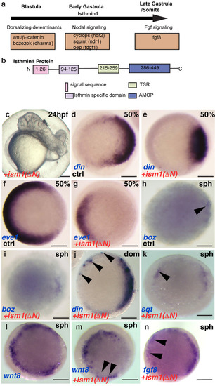
Dorsalization of embryos in ism1 deletion mutants. a Schematic representation of upstream regulators of ism1 during various stages of embryonic development. b Schematic representation of the Ism1 protein and the amino acid length of its domains, signal sequence, Isthmin specific domain (ISD), thrombospondin-type 1 repeat (TSR), and an adhesion-associated domain in Muc4 and other proteins (AMOP). c Dorsalized and cyclopic phenotype observed in ism1 (∆N) mRNA-injected embryos at 24 hpf (see Table 1). dg Dorsalization of the injected embryos at 50% epiboly, or late blastula (see Table 2). d, f Uninjected controls and e, g ism1(∆N)-injected embryos were probed for din and eve1 as dorsal and ventral markers, respectively. h, iboz appears unchanged after injection of ism1 (∆N). j, k, ndin, sqt, and fgf8 can be induced by ism1 (∆N) ectopically (arrowheads); l, m wnt8 (ORF1 + ORF2) is downregulated in the marginal zone. Left is ventral, and right is dorsal in all pictures. dn Animal pole views. The time of color development is shortened in k to emphasize the ectopic up-regulation of sqt expression. Scale bar: 200 μm

Expression Data
Genes:
Fish:
Anatomical Terms:
Stage: 50%-epiboly

Expression Detail
Antibody Labeling
Phenotype Data

Phenotype Detail
Acknowledgments
This image is the copyrighted work of the attributed author or publisher, and ZFIN has permission only to display this image to its users. Additional permissions should be obtained from the applicable author or publisher of the image. Full text @ Cell Tissue Res.