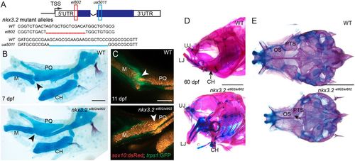
nkx3.2 mutant zebrafish are adult viable and develop craniofacial skeletal abnormalities. (A) Schematic of nkx3.2 mutant alleles: el802 – 14 bp deleted sequence including the ‘A’ of the ATG at the nkx3.2 translation start codon; ua5011 – 20 bp deleted sequence at the start of the homeobox DNA-binding domain. Coding regions of exons shown in blue and non-coding regions in white. (B) Lateral views of pharyngeal skeletal preparations stained with Alcian Blue (cartilage) and Alizarin Red (bone) show fusions of the jaw joint (arrowheads) in nkx3.2 mutants at 7 dpf. (C) Live imaging of transgenic juvenile zebrafish demonstrates loss of sox10:DsRed+/trps1:GFP+ jaw joint chondrocytes (arrowheads) in nkx3.2 mutants at 11 dpf. (D,E) Lateral views of the facial skeleton (D) and ventral views of the neurocranium (E) in adult nkx3.2 mutants at 60 dpf show craniofacial skeletal abnormalities including gaping jaw, fused jaw joint and displaced ceratohyal, and fusion of neurocranial orbitosphenoid and pterosphenoid bones (arrow in E). CH, ceratohyal; LJ, lower jaw; M, Meckel's cartilage; OS, orbitosphenoid; PQ, palatoquadrate; PTS, pterosphenoid; UJ, upper jaw; WT, wild type. Scale bars: 100 µm (B,C); 2 mm (D,E).
|
