ZFIN ID: ZDB-FIG-210305-4
Bastos de Oliveira et al., 2021 - Vasohibin-1 selectively regulates secondary sprouting and lymphangiogenesis in the zebrafish trunk. Development (Cambridge, England)   148(4) Full text @ Development
ADDITIONAL FIGURES
EXPRESSION / LABELING:
Genes:
Antibodies:
Fish:
Knockdown Reagent:
Anatomical Terms:
Stage Range: Prim-15 to Pec-fin
PHENOTYPE:
Fish:
Knockdown Reagent:
Observed In:
Stage Range: Prim-15 to Pec-fin

Fig. 4.

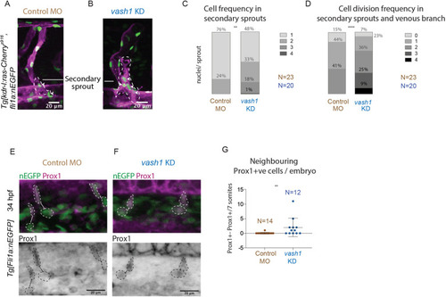
Secondary sprouts in vash1 morphants exhibit more Prox1+ cells and higher proliferation rates. (A,B) Secondary sprouts in Tg[kdr-l:ras-Cherrys916,fli1a:nEGFPy7] embryos, with membrane- and nuclei-labelled endothelial cells (EC) in control (A) and vash1 KD embryos (B). White arrowheads indicate nuclei in secondary sprouts (outlined by dashed line). (C,D) Quantification of the number of endothelial nEGFP-labelled nuclei in secondary sprouts immediately before connection to the ISV (C), and cell division frequency in the secondary sprout before and after connecting to the ISV, from 30 to 70 hpf (D). Quantifications are from three biological replicates. **P<0.0021, ****P<0.0002 (Mann–Whitney test in C, t-test in D). (E,F) Prox1-positive (Prox1+ve) EC were identified by immunostaining in Tg[fli1a:nEGFPy7] embryos in control and vash1 morphants. In controls, migrating cells in secondary sprouts are Prox1+ve and their neighbouring EC in the PCV are Prox1-negative (E). In vash1 morphants, both EC in the secondary sprout and the neighbouring EC still in the PCV are Prox1+ve (F). Analysed EC are highlighted (dashed line), and neighbouring EC are connected with a line. (G) Quantification of incidence of nEGFP+ Prox1+ve neighbouring EC per seven somites per embryo in both control and vash1 MO-injected embryos. **P<0.0021 (Mann–Whitney test). Pictures are representative of three replicated experiments.

Gene Expression Details
Gene Antibody Fish Conditions Stage Anatomy Assay
EGFP s916Tg; y7Tg control Pec-fin endothelial cell nucleus IFL
s916Tg; y7Tg + MO1-vash1 control Pec-fin endothelial cell nucleus IFL
mCherry s916Tg; y7Tg control Pec-fin endothelial cell membrane IFL
s916Tg; y7Tg + MO1-vash1 control Pec-fin endothelial cell membrane IFL
Antibody Labeling Details
Antibody Assay Fish Conditions Stage Anatomy
Ab5-prox1 IHC y7Tg control Prim-15 posterior cardinal vein angiogenic sprout
IHC y7Tg + MO1-vash1 control Prim-15 posterior cardinal vein angiogenic sprout
IHC Prim-15 posterior cardinal vein endothelial cell
Ab7-GFP IHC y7Tg control Prim-15 posterior cardinal vein endothelial cell
IHC y7Tg + MO1-vash1 control Prim-15 posterior cardinal vein endothelial cell
Phenotype Details
Fish Conditions Stage Phenotype
s916Tg; y7Tg + MO1-vash1 control Pec-fin angiogenic sprout cell division increased process quality, abnormal
Pec-fin angiogenic sprout cell population proliferation increased process quality, abnormal
Pec-fin angiogenic sprout endothelial cell increased amount, abnormal
y7Tg + MO1-vash1 control Prim-15 posterior cardinal vein endothelial cell ab5-prox1 labeling mislocalised, abnormal
Acknowledgments:
This image is the copyrighted work of the attributed author or publisher, and ZFIN has permission only to display this image to its users. Additional permissions should be obtained from the applicable author or publisher of the image. Full text @ Development