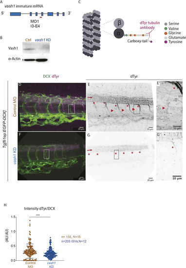
Endothelial microtubules are detyrosinated by Vash1 in zebrafish. (A,B) Knockdown (KD) strategy using a morpholino (MO) targeting the intron3-exon4 (I3-E4) (A) efficiently decreases Vash1 protein levels as detected by western blot (B). 48 hpf embryo lysate was used, three replicates were performed. (C) Mechanism of tyrosine cleavage from α-tubulin carboxy-terminus by Vash1, resulting in detyrosinated tubulin (dTyr). Detyrosination exposes a glutamate residue accessible by a custom-made antibody (Liao et al., 2019). (D-G′) Immunostainings of 48 hpf Tg[fli1ep:EGFP-DCX] embryos detect detyrosinated microtubules (referred to as dTyr; D-G) and GFP-labelled microtubules (referred as DCX; D,F). G and G′ show immunostaining of dTyr upon vash1 KD, compared with the control MO injected sibling embryos (E,E′). Arrows (E,G) indicate neural tube, with typically detyrosinated microtubules (E), reduced upon vash1 KD (G). Arrowheads indicate endothelial detyrosinated microtubules, only present in control embryos (E,E′). Asterisks (E,G) indicate motoneurons exhibiting high dTyr signal in control embryos (E,E′) and decreased dTyr signal in vash1 KD embryos (G,G′). Pictures are representative of three biological replicates. (H) Quantification of ratios between dTyr and DCX intensity signals of each ISV in control and vash1 KD groups. AU, arbitrary units. Each data point is one ISV, n=155 ISVs from 15 embryos (control) and n=205 ISVs from 12 embryos (vash1 KD), from three replicates. Data are mean±s.d. ***P<0.0002 (Mann–Whitney test). Pictures are representative of three replicated experiments.
|
