|
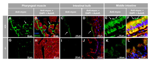
Immunohistochemistry of myocilin in non-ocular tissues of adult zebrafish. Wild-type and myoc KO adult (7 months) zebrafish tissue sections (14 μm) were incubated with a chicken anti-myocilin (TNT) primary antibody and a Cy2-conjugate goat anti-chicken IgY secondary antibody. Arrows show immunostaining in the periphery of pharyngeal muscular fibres (A,B,G,H), the enterocyte apical side in the intestinal bulb (C,D,I,J) and in the brush border of the epithelial cells in the middle intestine (E,F,K,L). Red signals correspond to tissue autofluorescence. bb: brush border. The images are representative of the results observed in three tissue sections from three animals. +/+: wild-type; −/−: myoc KO. Negative controls are shown in Figure S5.
|