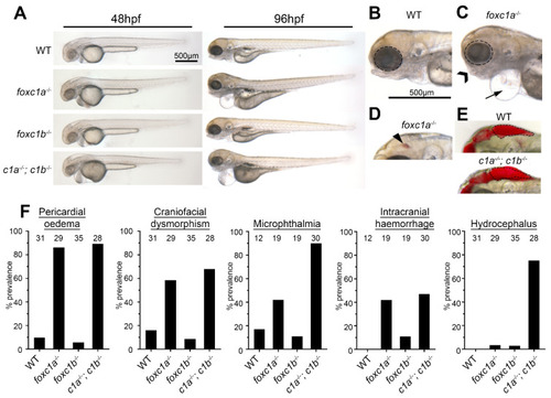
Foxc1 mutants exhibit multiple developmental defects. (A) foxc1 single mutants are largely indistinguishable from WT (wildtype) controls at 48 hpf (left panels), whereas foxc1a−/−; foxc1b−/− double homozygotes display hydrocephalus and oedema. By 96 hpf, no changes are observed in foxc1b−/− homozygotes, however; foxc1a−/− homozygotes and foxc1a−/−; foxc1b−/− double homozygotes display pericardial oedema (compare B and C, as highlighted by the arrow), microphthalmia (dotted circle), and craniofacial dysmorphism (chevron). At this stage, a subset of mutants also displayed intracranial hemorrhage (arrowhead in panel D), with foxc1a−/− homozygotes having greater frequency than foxc1b−/− homozygotes (42% vs. 11%, respectively), and (E) foxc1a−/−; foxc1b−/− double homozygotes present with hydrocephalus. (F) Quantification reveals that these defects are incompletely penetrant and generally more prevalent in double than single homozygotes (the number of embryos analyzed is shown above each bar). In the case of hydrocephalus, only the double homozygotes display an appreciable frequency of this phenotype.
|
