
Figure 3
- ID
- ZDB-FIG-210221-13
- Publication
- Pellegrini et al., 2020 - Induction of ER Stress in Acute Lymphoblastic Leukemia Cells by the Deubiquitinase Inhibitor VLX1570
- Other Figures
- All Figure Page
- Back to All Figure Page
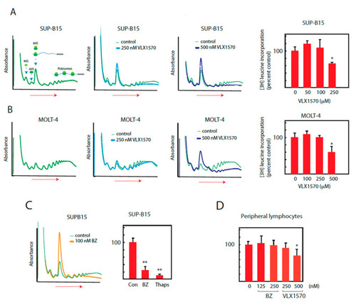
Suppression of translation by bortezomib and VLX1570. Cells were exposed to bortezomib or VLX1570, and ribosomes and polysomes were fractionated using sucrose gradient centrifugation. Incorporation of [3H]-leucine into acid precipitable material was measured after 6 h of drug exposure. (A,B) SUP-B15 or MOLT-4 cells were exposed to VLX1570 for 6 h and lysates were prepared in the presence of RNAase inhibitors and cycloheximide (100 μg/mL) and fractionated by sucrose gradient sedimentation (left to right). Absorbance (A280) was monitored during collection. The dose response of [3H]-leucine incorporation into acid precipitable material was determined after 6 h of drug exposure ([3H]-leucine was added during the last hour). Mean values ± S.D., * p < 0.05 Student′s t-test. (C) SUP-B15 cells were exposed to bortezomib (BZ) and lysates were fractionated by sucrose gradient sedimentation (left to right) and A280 monitored during collection. [3H]-leucine incorporation into acid precipitable material was measured after 6 h exposure to 100 nM bortezomib or 10 µM thapsigargin. Mean values + S.D., ** p < 0.01, Student′s t-test. (D) Peripheral lymphocytes were exposed to VLX1570 for 6 h and [3H]-leucine incorporation into acid precipitable material was determined. Mean values ± S.D., * p < 0.05, Student′s t-test. |