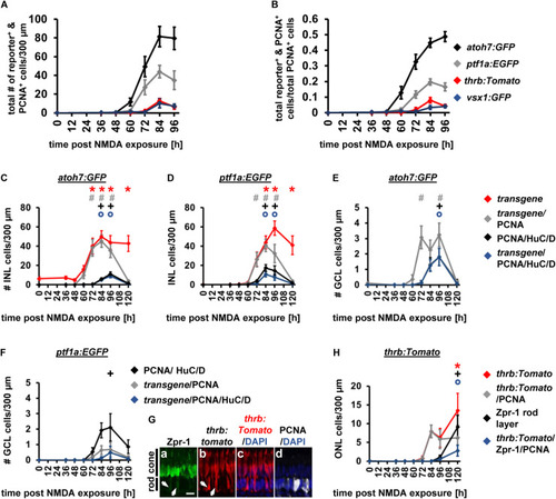
Comparison of the temporal expression patterns of neuronal competence factors in the NMDA-damaged retina. (A,B) Total number (ONL, INL, and GCL combined) of atoh7:GFP-, ptf1a: EGFP-, thrb:Tomato- and vsx1:GFP-positive cells (i.e., reporter-positive cells) that express PCNA (A) and when normalized to the total number of PCNA-positive cells in NMDA-damaged retinas (B). Mean ± SE, n ≥ 7. (C,D) Number of transgene-positive cells, transgene-positive cells that express PCNA and those that are triple-positive for the transgene, PCNA and HuC/D as well as the number of HuC/D and PCNA-positive cells in the INL of NMDA-exposed Tg[atoh7:GFP]rw021(C) and Tg[ptf1a:EGFP]jh1 zebrafish retinas [(D); 0, 36, 48, 60, 72, 84, 96, and 120 h post NMDA exposure]. (E,F) Number of transgene-positive cells that express PCNA and those that also co-localize with HuC/D as well as the number of HuC/D and PCNA-positive cells in the GCL of NMDA-exposed Tg[atoh7:GFP]rw021(E) and Tg[ptf1a:EGFP]jh1 zebrafish retinas (F). Mean ± SE, n ≥ 9. (G) Single confocal z-stack images of Tg[thrb:tomato]q22 retinas (Gb,c) at 120 h post NMDA exposure labeled for Zpr-1 (Ga), PCNA (Gd), and DAPI (Gc,d). Arrows, Zpr-1 and thrb:tomato-double positive cells in the rod photoreceptor cell nuclear layer. Scale bar, 10 μm (Ga). (H) Number of Zpr-1-positive cells, thrb:Tomato-positive cells, those that co-labeled with PCNA or those triple-positive for thrb:Tomato, PCNA and Zpr-1 in the rod ONL of NMDA-exposed retinas. Mean ± SE, n ≥ 8, *pTukey < 0.05, #p < 0.05, +p < 0.05, and °p < 0.05 indicate comparisons to 0 h post NMDA for the different measures that were assessed. The symbols are color-coded accordingto the line that they represent in the corresponding graphs (pANOVA, see Table 1).
|
