
Fig. 4
- ID
- ZDB-FIG-210218-59
- Publication
- Kim et al., 2020 - Notch Signaling Controls Oligodendrocyte Regeneration in the Injured Telencephalon of Adult Zebrafish
- Other Figures
- All Figure Page
- Back to All Figure Page
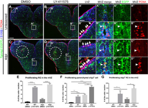
Fig. 4. Notch signaling is involved in the proliferation of olig2+ RG and parenchymal OPCs. All panels show transverse sections of the telencephalon, with the dorsal side at the top. (A~D”) Labeling of the telencephalon of Tg(olig2:EGFP) adult zebrafish with anti-S100β and -PCNA antibodies. (A, B) Treatment of wildtypes with DMSO (A) and LY-411575 to inhibit Notch signaling (B). Boxed areas indicate the LVZ (A’, B’) and MVZ (A”, B”). Arrows indicate PCNA+/S100β+ proliferating RG (B’), and the arrowhead indicates PCNA+/S100β+/olig2:EGFP+ proliferating RGs (B”). (C, D) Treatment of the injured telencephalon 4 dpl with DMSO (C) and LY-411575 to inhibit Notch signaling (D). Arrows indicate PCNA+/S100β+ proliferating RGs (C’, D’), and arrowheads indicate PCNA+/S100β+/olig2:EGFP+ proliferating RGs (C”, D”). (E~G) Quantification of proliferating RGs (S100β+/EGFP+/PCNA+) in the medial ventricular zone (E), proliferating parenchymal OPCs (S100β-/EGFP+/PCNA+) (F), and proliferating olig2+ RGs (S100β+/EGFP+/PCNA+) in the medial ventricular zone (G) (n=9 sections from two zebrafish, Mann–Whitney U test, *p<0.05, **p<0.01, ***p<0.001 and ****p<0.0001). Scale bars: A~D, 100 μm; A’~D”, 25 μm. DMSO, dimethyl sulfoxide; EGFP, enhanced green fluorescent protein; LVZ, lateral ventricular zone; MVZ, medial ventricular zone; OPCs, oligodendrocyte progenitor cells; PCNA, proliferating cell nuclear antigen; RG, radial glia. |