
Fig. 1
- ID
- ZDB-FIG-210218-55
- Publication
- Kim et al., 2020 - Notch Signaling Controls Oligodendrocyte Regeneration in the Injured Telencephalon of Adult Zebrafish
- Other Figures
- All Figure Page
- Back to All Figure Page
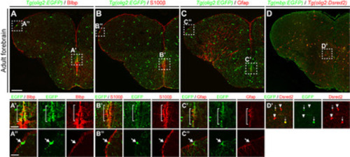
Fig. 1. olig2+ RG in the telencephalic ventricular zone of adult zebrafish. All panels show transverse sections of the telencephalon of adult zebrafish, with the dorsal side at the top. (A~C) Labeling of Tg(olig2:EGFP) zebrafish with various RG markers: anti-BLBP, -S100β, and -GFAP antibodies. Boxed areas indicate the MVZ (A’~C’) and LVZ (A”~C”) of the telencephalon. The bracketed area in (A’~C’) indicates the MVZ in which olig2+ RG exist. (A”~C”) arrows indicate olig2+ non-RGs in the LVZ. Seven zebrafish brains were analyzed in each indicated group and representative images were presented. (D~D’) Identification of mature oligodendrocytes in the telencephalic parenchymal region of the adult Tg(olig2:Dsred2)/Tg(mbp:EGFP) zebrafish. The boxed area in (D’) indicates the parenchymal region showing oligodendrocyte lineage cells. Arrows indicate mbp:EGFP+/olig2:Dsred+ mature oligodendrocytes, and arrowheads indicate mbp:EGFP-/olig2:Dsred+ OPCs (n=8 sections from one zebrafish). Scale bars: A~D, 100 μm; A’~C”, 25 μm. GFAP, glial fibrillary acidic protein; EGFP, enhanced green fluorescent protein; LVZ, lateral ventricular zone; MVZ, medial ventricular zone; OPCs, oligodendrocyte progenitor cells; RG, radial glia. |