
Fig. 7
- ID
- ZDB-FIG-210217-77
- Publication
- Robles et al., 2020 - The zebrafish visual system transmits dimming information via multiple segregated pathways
- Other Figures
- All Figure Page
- Back to All Figure Page
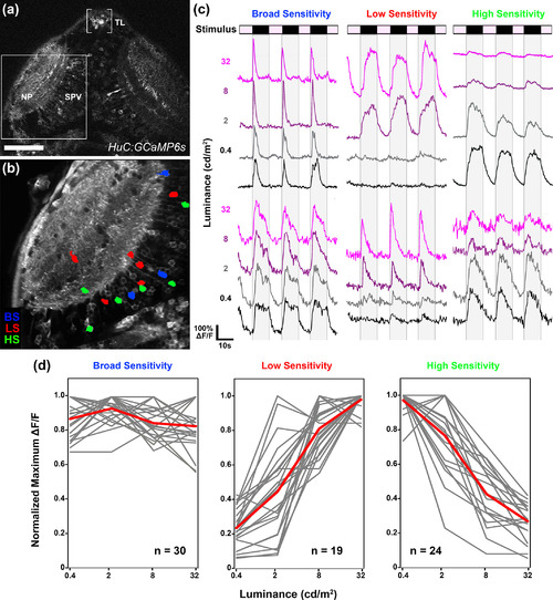
Optic tectum contains dimming‐responsive neurons with different photosensitivities. (a) Low‐magnification dorsal view of a 6 dpf Tg(Elavl3:gal4,uas:gcamp6s) larva. Boxed region indicates typical field of view used for functional imaging trials. (b) Average intensity projection from a recording in the left tectum of a 6 dpf Tg(elavl3:gal4,uas:gcamp6s) larva presented with light steps to the right eye. Region of interest color coding indicates photosensitivity profile of dimming‐responsive tectal neurons: broad sensitivity (BS; blue), low sensitivity (LS; red), and high sensitivity (HS; green). Note sparse and dispersed distributions of neurons in both the neuropil (NP) and periventricular (SPV) layers of the tectum. (c) Sample traces of individual BS, LS, and HS tectal neurons. Each trace is 58 s in duration and consists of four ON phases of 7‐s duration interleaved with three OFF phases of 10‐s duration. (d) Photosensitivity tuning curves for 73 dimming‐responsive neurons (BS, LS, and HS) detected in 10 tecta (gray). Average of individual traces is shown in red. Scale bar: 80 μm in (a), 40 μm in (b) |