
Fig. 2
- ID
- ZDB-FIG-210217-72
- Publication
- Robles et al., 2020 - The zebrafish visual system transmits dimming information via multiple segregated pathways
- Other Figures
- All Figure Page
- Back to All Figure Page
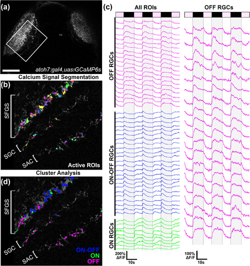
Functional identification of OFF retinal ganglion cell (OFF‐RGC) terminals in vivo. (a) Low‐magnification dorsal view of a 6 dpf Tg(atoh7:gal4,uas:gcamp6s) larva. Boxed region indicates typical field of view used for functional imaging trials. (b) Average intensity projection from a recording in the left tectum of a Tg(atoh7:gal4,uas:gcamp6s) larva presented with dimming stimuli to the right eye. Active regions of interest (ROIs) identified by cross‐correlation‐based image segmentation are displayed in different colors. Note multiple active regions in the SAC/SPV, SGC, and deepest sublayers of SFGS. (c) Fluorescence intensity traces of ROIs indicated in C during repeated presentation of dimming steps to the contralateral eye as indicated by light and dark bars at top. Each trace is 58 s in duration and consists of ON phases of 7‐s duration and OFF phases of 10‐s duration. Gray shading overlaid on traces indicates dimming phase when stimulus display is OFF. k‐means cluster analysis divided ROIs into three groups based on their temporal dynamics, indicated by color coding in blue, green, and magenta. Magenta‐colored traces represent dimming detector RGCs that exhibited strong and consistent OFF responses. (d) Average intensity projection shown in (b) presented with OFF ROI color coding that corresponds to clustering results shown in (c). Note that in this larva OFF‐RGCs ROIs are located in the SAC/SPV, SGC, and deepest sublayers of SFGS (5/6). Data are from 52 ROIs detected in a single larva. Scale bar: 50 μm in (a), 20 μm in (b,d). SAC, stratum album centrale; SFGS, stratum fiibrosum et griseum superficiale; SGC, stratum griseum centrale; SPV, stratum periventriculare |