
Fig. 5
- ID
- ZDB-FIG-210210-70
- Publication
- Bürgi et al., 2020 - Ligand Binding to the Collagen VI Receptor Triggers a Talin-to-RhoA Switch that Regulates Receptor Endocytosis
- Other Figures
- All Figure Page
- Back to All Figure Page
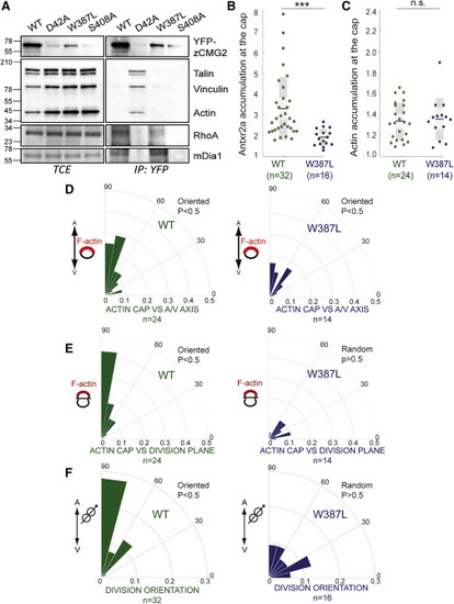
Figure 5. Loss of zAntrx2’s Ability to Bind Talin Affects Oriented Epiblast Cell Division (A) Zebrafish embryos were injected with mRNA of WT, W387L, or S408A zANtxr2a-eYFP, lysed, and zANtxr2a was immunoprecipitated followed by SDS-PAGE and western blotting against talin, vinculin, actin, RhoA, mDia, and GFP. (B) Antxr2a accumulation at the cap in epiblast cells. Zebrafish embryos were injected with either WT-Antxr2a-eYFP or W387L-Antxr2a-eYFP. The fluorescence intensity of the WT or the W387L fluorescently tagged versions of Antxr2a was then measured along the plasma membrane of epiblast cells. The intensity profile was fitted to a Gaussian distribution and the accumulation of the fluorescence intensity was calculated as described in the STAR Methods. (C) F-actin accumulation at the cap in epiblast cells. Zebrafish embryos were injected with LifeAct-mRFP biosensor and with either WT or W387L-Antxr2a-eYFP. The fluorescence intensity accumulation of the LifeAct-mRFP biosensor in one side of the cell was measured as described in (B). (B and C) For statistical analysis, an unpaired t test was performed comparing the WT to the W387L zAntrx2a mutants, ∗∗∗p < 0.001; n.s: p > 0.05 (D) Polar graphs showing the frequency distribution of the angles between the positions of the actin F-cap and the embryonic axis (A-V axis) in embryos expressing WT (green) or W387L (blue)-Antxr2a-eYFP. In all cases, the F-actin cap is aligned with the embryonic axis. (E) Polar graphs showing the frequency distribution of the angles between the F-actin cap and the division plane. Although in WT-Antxr2a-eYFP expressing embryos (green), there is a correlation between the F-actin cap and the division plane, in embryos expressing W387L-Antxr2a-eYFP (blue) this correlation is lost. (F) Polar graphs showing the frequency distribution of angles of division of epiblast cells. While divisions are oriented along the embryonic axis in embryos expressing WT-Antxr2a-eYFP (green), divisions are randomized in epiblast cells of embryos expression either W387L (blue). |
Reprinted from Developmental Cell, 53, Bürgi, J., Abrami, L., Castanon, I., Abriata, L.A., Kunz, B., Yan, S.E., Lera, M., Unger, S., Superti-Furga, A., Peraro, M.D., Gaitan, M.G., van der Goot, F.G., Ligand Binding to the Collagen VI Receptor Triggers a Talin-to-RhoA Switch that Regulates Receptor Endocytosis, 418-430.e4, Copyright (2020) with permission from Elsevier. Full text @ Dev. Cell