
Fig. 4
- ID
- ZDB-FIG-210129-25
- Publication
- Ghilardi et al., 2020 - Expression pattern of the small muscle protein, X-linked (smpx) gene during zebrafish embryonic and larval developmental stages
- Other Figures
- All Figure Page
- Back to All Figure Page
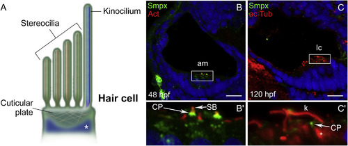
Smpx localizes to the cuticular plate of the inner ear hair cells. (A) Schematic representation of the hair cell apical membrane of the zebrafish inner ear; the kinocilium, with the structural tubulin organized in microtubules (in blue), and the bundle of F-actin-based mechanosensitive stereocilia (in brown) are depicted. The cuticular plate and the somatic tubulin (white asterisk) are also shown. (B,C) Paraffin sections co-labeled with antibodies against Smpx and phalloidin (B) and with antibodies against Smpx and acetylated tubulin (C), with the nuclei stained with DAPI. (B′,C′) 5X magnifications of B and C; Smpx is located in the region corresponding to the cuticular plate, in between the stereociliary F-actin-based bundle above (B′) and the somatic tubulin below (C′); F-actin (red) and Smpx (green) signals co-localize in the uppermost part of the cuticular plate (yellow in B′). Images are all lateral views, anterior to the left, of confocal Z-stacks taken from paraffin sections from embryos and larvae. am, anterior macula; lc, lateral crista; CP, cuticular plate; SB, stereociliary bundle; k, kinocilium. Scale bars = 30 μm. |
| Gene: | |
|---|---|
| Antibody: | |
| Fish: | |
| Anatomical Term: | |
| Stage Range: | Long-pec to Day 5 |
Reprinted from Gene expression patterns : GEP, 36, Ghilardi, A., Diana, A., Prosperi, L., Del Giacco, L., Expression pattern of the small muscle protein, X-linked (smpx) gene during zebrafish embryonic and larval developmental stages, 119110, Copyright (2020) with permission from Elsevier. Full text @ Gene Expr. Patterns