
|
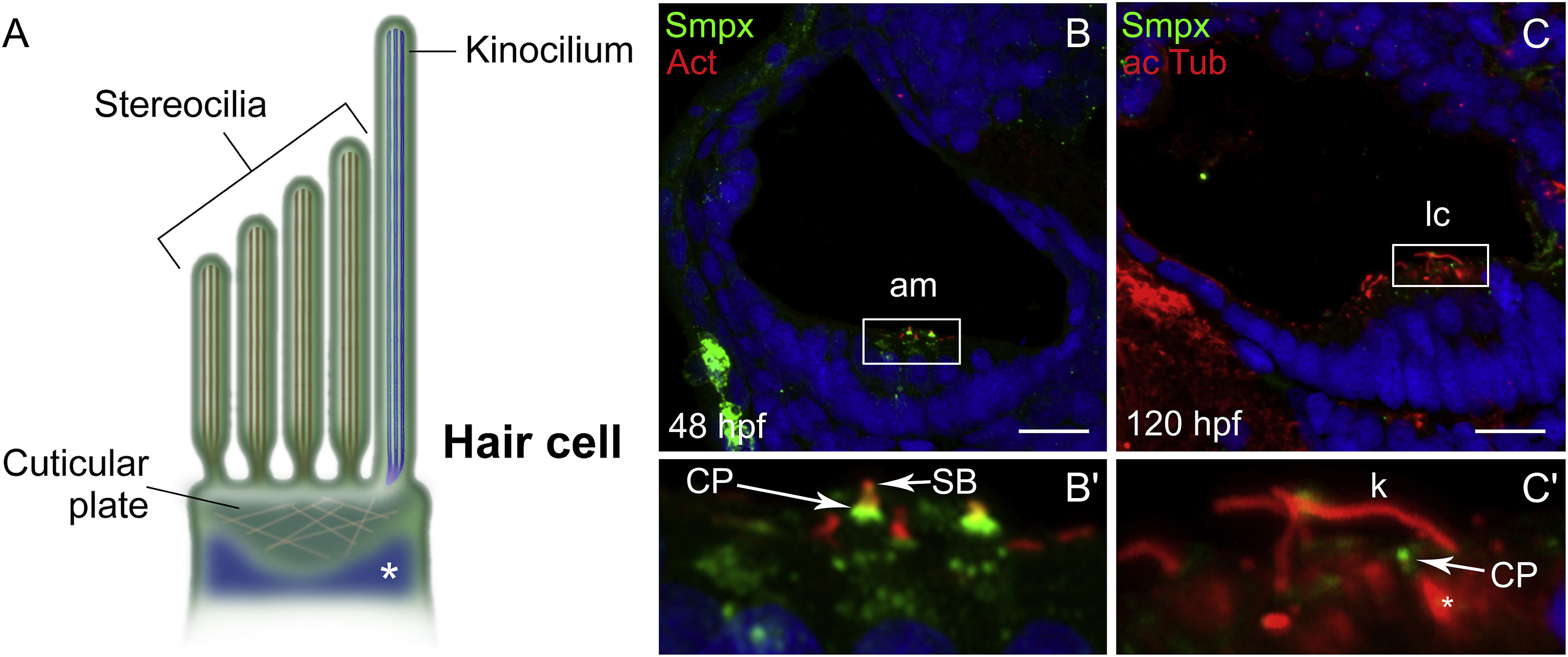
Fig. 4 Smpx localizes to the cuticular plate of the inner ear hair cells. (A) Schematic representation of the hair cell apical membrane of the zebrafish inner ear; the kinocilium, with the structural tubulin organized in microtubules (in blue), and the bundle of F-actin-based mechanosensitive stereocilia (in brown) are depicted. The cuticular plate and the somatic tubulin (white asterisk) are also shown. (B,C) Paraffin sections co-labeled with antibodies against Smpx and phalloidin (B) and with antibodies against Smpx and acetylated tubulin (C), with the nuclei stained with DAPI. (B′,C′) 5X magnifications of B and C; Smpx is located in the region corresponding to the cuticular plate, in between the stereociliary F-actin-based bundle above (B′) and the somatic tubulin below (C′); F-actin (red) and Smpx (green) signals co-localize in the uppermost part of the cuticular plate (yellow in B′). Images are all lateral views, anterior to the left, of confocal Z-stacks taken from paraffin sections from embryos and larvae. am, anterior macula; lc, lateral crista; CP, cuticular plate; SB, stereociliary bundle; k, kinocilium. Scale bars = 30 μm.
Reprinted from Gene expression patterns : GEP, 36, Ghilardi, A., Diana, A., Prosperi, L., Del Giacco, L., Expression pattern of the small muscle protein, X-linked (smpx) gene during zebrafish embryonic and larval developmental stages, 119110, Copyright (2020) with permission from Elsevier. Full text @ Gene Expr. Patterns