
FIGURE 4
- ID
- ZDB-FIG-210128-19
- Publication
- Zhu et al., 2021 - Tmc Reliance Is Biased by the Hair Cell Subtype and Position Within the Ear
- Other Figures
- All Figure Page
- Back to All Figure Page
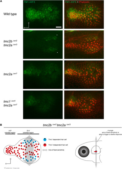
Hair cells of the posterior macula are differentially dependent on Tmc proteins. (A) Representative images of FM1-43FX (green) uptake in posterior maculae of wild-type and mutant larvae at 7 dpf. Hair bundles are labeled with phalloidin (red). In a wild-type macula, passage of FM1-43FX is observed in all hair cells. In the tmc2bcwr2 tmc2acwr3 double mutant, FM1-43FX uptake is limited to two symmetrical stripes of hair cells located toward the posterior of the macula at the dorsal and ventral poles (arrowheads). In contrast, the tmc2acwr7 single mutant or tmc1cwr4 tmc2acwr7 double mutant hair cells do not reveal a pattern similar to the tmc2bcwr2 tmc2acwr3 double mutant. A, anterior; D, dorsal. Scale bar = 10 μm. Similar patterns of saccular uptake were observed for five larvae for each mutant type and control. (B) (Left) Map of hair cell positions and hair bundle polarities in relation to Tmc1 use within the posterior macula based on the tmc2bcwr2 tmc2acwr3 double mutant. Each color circle (red or blue) represents an individual hair cell, and the black dot within represents the position of the kinocilium, which reflects bundle polarity. Double-headed arrow indicates axis of best sensitivity preserved in the tmc2bcwr2 tmc2acwr3 double mutant. (Right) Schematized zebrafish head depicting just one axis of best sensitivity (dorsal-ventral) able to elicit a startle response. |