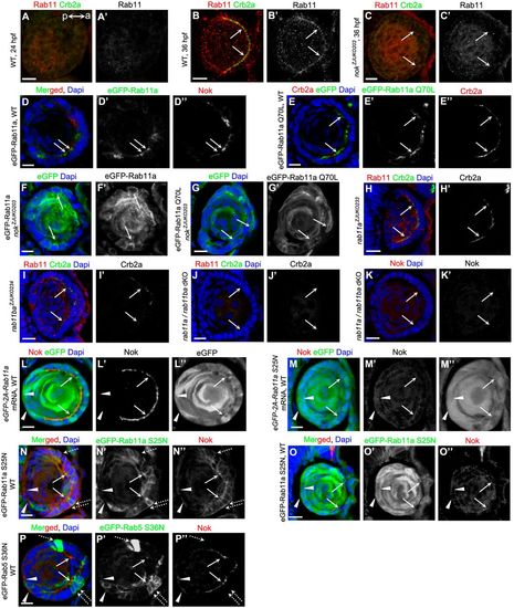
The apical localizations of Nok and Rab11 are reciprocally dependent. (A,B) Temporospatial expression pattern of Rab11 in zebrafish lens. Rab11 did not display polarized aggregation in epidermal cells when Crb2a was not detectable at 24 hpf (A), but strongly aggregated in the apical regions in lens epithelial cells at 36 hpf (B). a (anterior) and p (posterior) show the lens orientation. (C) Rab11 lost its apical enrichment in lens epithelial cells in nok mutants. (D,E) eGFP-tagged Rab11a (D) and Rab11 Q70L (E) proteins were enriched in the apical regions in lens epithelial cells in WT. Overexpression of eGFP-Rab11a and eGFP-Rab11 Q70L did not significantly affect the apical enrichment of Nok. (F,G) eGFP-tagged Rab11a (F) and Rab11 Q70L (G) proteins were distributed at the lateral and basal regions in lens epithelial cells in nok mutants. (H-K) The apical localization of Crb2a in lens epithelial cells was partially disrupted in rab11a (H) or rab11ba mutants (I) and completely disrupted in rab11a/rab11ba dKO mutants (J). Similarly, the apical localization of Nok was lost in rab11a/rab11ba dKO mutants (K). (L,M) Overexpression of eGFP-2A-Rab11a (L) or eGFP-2A-Rab11a S25N (M) following mRNA injection. Overexpression of Rab11a S25N (M), but not Rab11a (L), impeded the apical aggregation of Nok. Low levels of Nok were observed in mesenchymal cells overexpressing eGFP-2A-Rab11a S25N in the posterior lens (M, white triangles). (N,O) eGFP-Rab11a S25N is evenly spread to apical, lateral and basal regions in WT lens epithelial cells. Lower level overexpression of eGFP-Rab11a S25N induced the lateral and basal distribution of Nok (N), and higher level of eGFP-Rab11a S25N impeded the apical aggregation of Nok (O). Low levels of Nok were observed in mesenchymal cells overexpressing eGFP-Rab11a S25N in the posterior lens (white triangles). (P) Overexpression of eGFP-Rab5 S36N did not significantly affect the apical enrichment of Nok in WT lens epithelial cells. Low levels of Nok (white triangles) were observed in mesenchymal cells overexpressing eGFP-Rab5 S36N in the posterior lens. The embryos used for B-P were fixed at 36 hpf. Arrows show the apical regions of lens epithelial cells. Broken arrows show the basal regions of lens epithelial cells. White triangles show fiber cells in the posterior lens. Plasmids pTol2-EF1::eGFP-Rab11a, pTol2-EF1::eGFP-Rab11a S25N, pTol2-EF1::eGFP-Rab11a Q70L or pTol2-EF1::eGFP-Rab5 S36N were used for the sporadic overexpression of eGFP-tagged Rab proteins shown in Fig. 3D-G,N-P. Scale bars: 10 μm.
|
