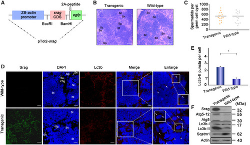
srag transgenic zebrafish analysis. (A) Schematic depiction of the pTol2-srag-egfp transgenic construct. The transgenic structure contains a region of the zebrafish actin promoter linked with the srag CDS (coding DNA sequence), a gfp tag, and 2A peptide cloned into the pTol2 vector. (B) Histological analysis of adult testis samples of srag transgenic and wild-type zebrafish by H&E staining. The white arrows indicate spermatogonia (sg) and Sertoli cells (sn). Scale bar: 50 μm. (C) Spermatid number in germ cell cyst in testis between srag transgenic and wild-type zebrafish (P < 0.9). (D) Immunofluorescence analysis of testis samples in both transgenic and wild-type zebrafish using anti-LC3B antibody. Images were captured using confocal microscopy. Lc3b-II puncta were obviously increased in the srag transgenic testis in comparison with wild type in spermatids (St), spermatocytes (Sc), and spermatogonia (Sg). The nuclei were stained by DAPI (blue). The enlarged images on the right panels originated from the white squares, the white arrows indicate Lc3-II puncta in spermatocytes (Sc). Enlarged box within the image highlights Lc3b-II puncta in Sertoli cells (Sn). Scale bar: 10 μm. (E) The number of Lc3b-II puncta was quantified from ∼20 cells for each group from (D). Data were presented as means ± SD. T-test was performed, *P < 0.05; **P < 0.01 (n = 3 independent experiments). (F) Western blot analysis of testis samples in both srag transgenic and wild-type zebrafish. Srag protein was detected in transgenic testis but not in wild type using the anti-Srag antibody. β-Actin was used as a control. Western blot analysis indicated that Lc3b-II and Atg5-Atg12 were upregulated, whereas Sqstm1 was downregulated in testis of transgenic zebrafish.
|
