FIGURE

Figure 2

ID
ZDB-FIG-201209-17
Publication
Terzioglu et al., 2020 - Improving CRISPR/Cas9 mutagenesis efficiency by delaying the early development of zebrafish embryos
Other Figures
All Figure Page
Back to All Figure Page
Figure 2

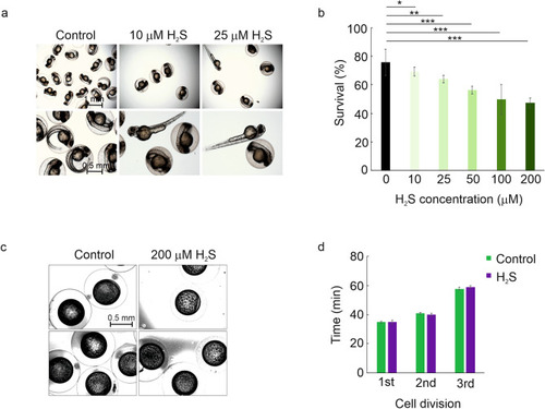
The effect of H2S treatment on the early development of zebrafish embryos. (a,b) H2S concentrations of 10–25 µM cause no changes in the morphology and minor decrease in the survival of zebrafish embryos, while the concentrations of 50–200 µM cause substantial increase in both the rate of malformation and mortality. Fertilized eggs were incubated in E3 water supplemented with 10–200 µM H2S for 48 h. The survival and well-being of the embryos was followed for 48 h and compared to E3 treated group (control). n(Control) = 397, n(10 µM) = 358, n(25 µM) = 310, n(50 µM) = 130, n(100 µM) = 182, n(200 µM) = 208. (c) Comparative developmental stages of control (E3 treated) and H2S treated embryos. Newly fertilized eggs were kept in E3 water with 200 µM H2S and the development was followed by time-lapse imaging. Two representative images are shown for both treatments (control images on the left, H2S treatment images on the right). (d) The times for the first, the second and the third cell division in H2S treated and control (E3 treated) embryos. Error bars represent SD, *p < 0.05, **p < 0.01, ***p < 0.001.

Expression Data

Expression Detail
Antibody Labeling
Phenotype Data

Phenotype Detail
Acknowledgments
This image is the copyrighted work of the attributed author or publisher, and ZFIN has permission only to display this image to its users. Additional permissions should be obtained from the applicable author or publisher of the image. Full text @ Sci. Rep.