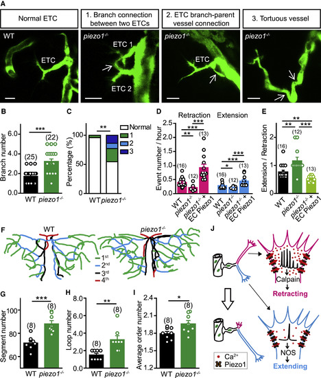
Zebrafish piezo1 Mutation Impairs ETC Pathfinding and Brain Vascular Patterning (A) Images of ETC morphology in 3-dpf Tg(kdrl:eGFP) larvae with WT or piezo1−/− background. The white arrows indicate abnormal connections. Scale bars, 10 μm. (B) Summary of the branch number of ETCs in WT siblings and piezo1−/− larvae. (C) Percentages of normal and abnormal morphologies of ETCs and vessels. WT, from 25 ETCs in 12 larvae; piezo1−/−, from 22 ETCs in 8 larvae. (D and E) Summary of the retraction and extension (D) and extending/retracting ratio (E) of ETC branches in WT, piezo1−/−, and piezo1−/−+EC Piezo1. WT siblings, from 16 ETCs in 9 larvae; piezo1−/−, from 12 ETCs in 6 larvae; piezo1−/−+EC Piezo1, from 13 ETCs in 7 larvae. (F–I) Representative skeletons (F) and summary for the vessel segment number (G), internal vessel loop number (H), and average order number of vessel segments (I) of the midbrain vasculature in WT siblings and piezo1−/− larvae at 7 dpf. The numbers in the brackets represent the numbers of larvae examined. (J) Working model. The same datasets were used in (B) and (C) or in (D) and (E). ∗p < 0.05; ∗∗p < 0.01; ∗∗∗p < 0.001 (two-tailed Mann-Whitney test for B, E, and H; Fisher exact test for C; and unpaired two-tailed Student’s t test for D, G, and I). Mean ± SEM. See also Figure S6.
|
