
FIGURE
Figure 3—figure supplement 3—source data 1.
- ID
- ZDB-FIG-201003-88
- Publication
- Sha et al., 2020 - Erasable labeling of neuronal activity using a reversible calcium marker
- Other Figures
- All Figure Page
- Back to All Figure Page
Figure 3—figure supplement 3—source data 1.
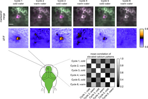
Top panels are individual Z slices from the pallium of the same fish (5 dpf) brain illustrating the same field of view from six marking cycles of alternating cold and warm water stimulus. Scale bar is 10 μm. Bottom-right panel is a normalized correlation matrix comparing labeling patterns in the upper pallium and habenula across multiple marking cycles in the same fish. |
Expression Data
Expression Detail
Antibody Labeling
Phenotype Data
Phenotype Detail
Acknowledgments
This image is the copyrighted work of the attributed author or publisher, and
ZFIN has permission only to display this image to its users.
Additional permissions should be obtained from the applicable author or publisher of the image.
Full text @ Elife