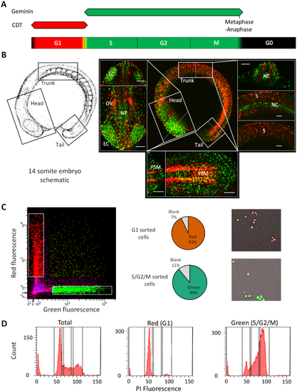
FACS mediated separation of cells by cell cycle stage in the developing embryo. (A) Schematic of fluorescence from the FUCCI system through the cell cycle with colours indicating phases marked by the fluorescent reporter genes. (B) Left panel; a schematic of the 14-somite embryos, reproduced with permission from (15) (© 1995 WILEY-LISS, INC.). Right panel; center: max projection fluorescent image of a 14-somite FUCCI transgenic embryo, showing the distribution of rapidly (green) and slowly cycling (red) cells. Surrounding panels: higher magnification views of the head, trunk and tail regions with alternate views shown in the top/right sections and cross-sections (denoted by the dashed lines) shown in the bottom/ left sections of each surrounding panel. Scale bar = 50 uM. Abbreviations, otic vesicle (OV), eye cup (EC), notochord (NC), somites (S), neural tube (NT), pre-somitic mesoderm (PSM). (C) FACS sorting of FUCCI embryos, from left to right, FACS plot showing gating, pie charts showing cell selection efficiency and representative fluorescent images of isolated cells. (D) Propidium Iodide DNA content analysis of isolated cells. FACS traces show a shift in isolated cell DNA content from a level in accordance with primarily Gap phase 1 cells (G1) (gated as P3) in the red population to principally S and G2 phase cells (gated in P4 and 5) in the green population, emanating from a mixed total population. Proportions are shown in Supplementary Table S1.
|
