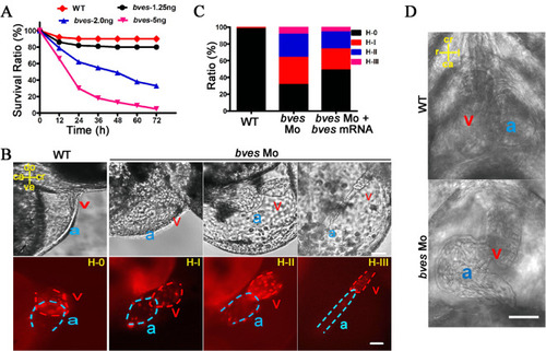
bves knockdown led to abnormal cardiac looping in zebrafish. (A) Survival ratio of wild type and morphants with 1.25, 2.0 and 5.0 ng of bves Mo at 72 hpf. WT, wild type; bves-1.25 ng, bves morphants with injection of 1.25 ng of bves Mo; bves-2.0 ng, bves morphants with injection of 2.0 ng of bves Mo; bves-5.0 ng, bves morphants with injection of 5.0 ng of bves Mo. (B) bves morphants showed cardiac defects at 48 hpf for the cmlc2:dsRed transgenic fish (a: atrium; v: ventricle), which were divided into four types based on the degree of cardiac dysplasia. H-0, the normal phenotypes; H-I, the moderate phenotypes; H-II, the strong phenotypes; H-III, the severe phenotypes. The position of the heart is marked by compass lines. ca, caudal; cr, cranial; do, dorsal; ve, ventral. Scale bar: 50 μm. (C) Data statistics of the different cardiac phenotypes in (B). (D) The morphants at 72 hpf with a left-looping heart in the ventral view. ca, caudal; cr, cranial; r, right; l, left. Scale bar: 50 μm. WT: wild type; bves Mo: bves morphants; bves Mo + bves mRNA, coinject bves morpholino and bves mRNA.
|
