FIGURE

Figure 2—figure supplement 2.

ID
ZDB-FIG-200822-38
Publication
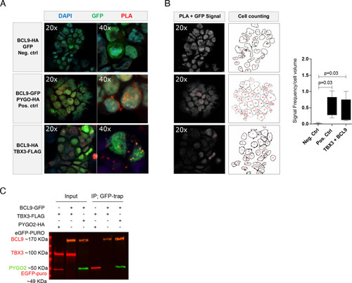
Zimmerli et al., 2020 - TBX3 acts as tissue-specific component of the Wnt/β-catenin transcriptional complex
Other Figures
All Figure Page
Back to All Figure Page
Figure 2—figure supplement 2.

Cytological proximity of BCL9 and TBX3.

(A) Proximity ligation assay (PLA) confirms vicinity between BCL9 and TBX3 when expressed in human HEK293T cells: red signal is observed upon protein-protein proximity. Nuclear GFP and BCL9 do not give rise to PLA signal (upper panel, negative control). PYGO2 is used as positive interactor with BCL9 (middle panel) and gives rise to PLA signal (red). TBX3 and BCL9 produce nuclear PLA signal in a subset of transfected cells (detected with GFP; lower panel). A representative image in 20x and 40x magnification and for each condition is shown (B) Quantification of the PLA. Signal of interest (GFP+PLA signal) was detected and normalized to the cell amount. A representative image of the quantification procedure for each condition is shown. The average of PLA quantification is presented as Tukey style box plot graph (at least three images per condition were analyzed). Significant p-values obtained with Mann–Whitney U test are shown. (C) Immunoprecipitation assay using a GFP-trap strategy. BCL9 reliably interacts with HA-tagged PYGO2 in high-salt washing conditions (400 mM NaCl). In this stringent condition, FLAG-tagged TBX3 is not observed when GFP-BCL9 is immunoprecipitated, suggesting absence of direct interaction or affinity inferior to that between BCL9 and PYGO2. IP of GFP is used as control.

Expression Data

Expression Detail
Antibody Labeling
Phenotype Data

Phenotype Detail
Acknowledgments
This image is the copyrighted work of the attributed author or publisher, and ZFIN has permission only to display this image to its users. Additional permissions should be obtained from the applicable author or publisher of the image. Full text @ Elife