
Fig. 3
- ID
- ZDB-FIG-200820-23
- Publication
- El-Nachef et al., 2020 - De novo enteric neurogenesis in post-embryonic zebrafish from Schwann cell precursors rather than resident cell types
- Other Figures
- All Figure Page
- Back to All Figure Page
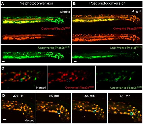
Enteric neurogenesis persists in the post-embryonic development despite the apparent absence of resident neuronal precursors. (A,B) 2D projection of z stack from a 4.5 dpf Phox2b-kaede fish demonstrates green fluorescent enteric neurons, but no red fluorescent cells (A). Yolk and intraluminal mucous exhibit expected autofluorescence in both channels. After photoconversion of all Phox2b-kaede neurons in the gut, all enteric neurons fluoresce red, although some retain decreased green fluorescence (B). (C) Live imaging 12 h after photoconversion at 4.5 dpf reveals the appearance of green fluorescent enteric neurons in the intestine with no red fluorescence (a representative neuron is circled), indicating that these neurons did not arise from pre-existing red fluorescent Phox2b-kaede cells. (D) Live 2D projection of a 10 h time-lapse after photoconversion at 4.5 dpf detects the emergence of de novo enteric neurons (circled), as indicated by the gradual appearance of a green-only neuron in a region of the intestine that was originally not occupied by a neuron. Scale bars: 20 µm. |