
Fig. 2
- ID
- ZDB-FIG-200818-16
- Publication
- Begovich et al., 2020 - Phosphoribosyl pyrophosphate synthetase polymerization influences lens fiber organization in zebrafish
- Other Figures
- All Figure Page
- Back to All Figure Page
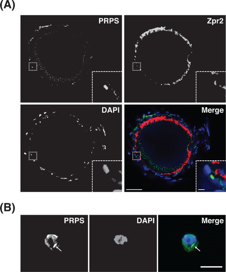
Phosphoribosyl pyrophosphate synthetase (PRPS) cytoplasmic filaments assemble in cells adjacent to the retinal pigmented epithelium (RPE). A, Single optical slice from a confocal z-stack shows a representative example of a 5 dpf eye stained for PRPS (green), Zpr2 RPE marker) (red), and 40,6-diamidino- 2-phenylindole (DAPI) (blue) and depicts PRPS filament formation in the cytoplasm of cells adjacent to the RPE layer. Rostral is to the left and dorsal is to the top. Insets in bottom right corners show zoomed views of boxed areas. Scale bars: 40 μm for main images, 3 μm for insets (n = 6 embryos). B, Representative maximum intensity projections of confocal images of a single cell, dissected from retinal tissue and stained for PRPS (green) and DAPI (blue), depict cytoplasmic localization of PRPS filaments. Retinas were dissected, subjected to trypsin treatment, and plated before being fixed and immunostained. Arrow indicates PRPS filament. Scale bar: 10 μm. (n = 3 eye dissections; ~30% of the dissociated cells exhibited PRPS filaments) |
| Antibody: | |
|---|---|
| Fish: | |
| Anatomical Term: | |
| Stage: | Day 5 |