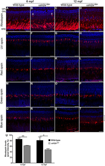
Rod and cone opsin localization in the ush2au507 retina. Rhodopsin (A–D) and UV (E–H), red (I–L), green (M–P) and blue (Q–T) cone opsins were detected in the wt and ush2au507 zebrafish retina at 6 months post fertilization (mpf), and 12 mpf by immunohistochemical analysis (red). In the 6 mpf ush2au507 retina, rhodopsin was partially mislocalized in some regions where it was detected in parts of the rod photoreceptors other than the outer segment (arrows in B). In the 12 mpf mutant retina, rhodopsin was more extensively mislocalized and expression in the rod outer segments was reduced compared to wt. Blue opsin was noted in the blue cone inner segments (highlighted with a bracket) in the ush2au507 retina at 12 mpf (T). All sections are counterstained with DAPI nuclei acid stain (blue). Bar chart (U) shows levels of rhodopsin detected in the rod outer segments at 6 and 12 mpf, measured using pixel intensity on confocal images (mean ± SEM. Three sections analyzed per fish, n = 3 per age). Unpaired t-tests were used to compare wt and ush2au507 rhodopsin levels at each age. *P < 0.05, **P < 0.01. OPL, outer plexiform layer; ONL, outer nuclear layer; RPE, retinal pigment epithelium. Scale bar = 25 μm.
|
