
Fig. 7
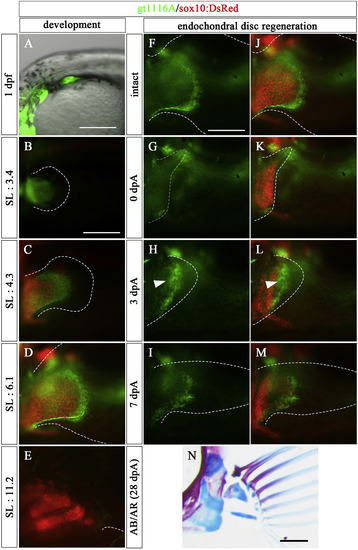
Expression patterns of gt1116A in pectoral fin development and Type 1 regeneration (5.0–7.0 mm SL larvae). (A–E) Expression pattern of UAS:EGFP in the pectoral fin of a gt1116A transgenic fish observed at embryonic (1 dpf) and post-hatching stages (SL: 3.4, 4.3, 6.1, and 11.2 mm) with observation of chondrocytes using sox10:DsRed. (F–N) The amputated pectoral fin at the middle of endochondral disc in a gt1116A transgenic fish. (F–I) Expression pattern of UAS:EGFP of gt1116A. (J–M) Merged images of expression pattern of the UAS:EGFP and sox10:DsRed. White arrowheads show re-expression of UAS:EGFP at 3 dpA. (N) the pectoral fin skeletons stained with AR and AB at 28 dpA. Dashed lines indicate the outline of fins and the UAS:EGFP-positive mesenchymal cells in the regenerated region, respectively. Scale bars: 200 μm in A, B, F, and N. |
Reprinted from Developmental Biology, 463(2), Yoshida, K., Kawakami, K., Abe, G., Tamura, K., Zebrafish can regenerate endoskeleton in larval pectoral fin but the regenerative ability declines, 110-123, Copyright (2020) with permission from Elsevier. Full text @ Dev. Biol.