
|
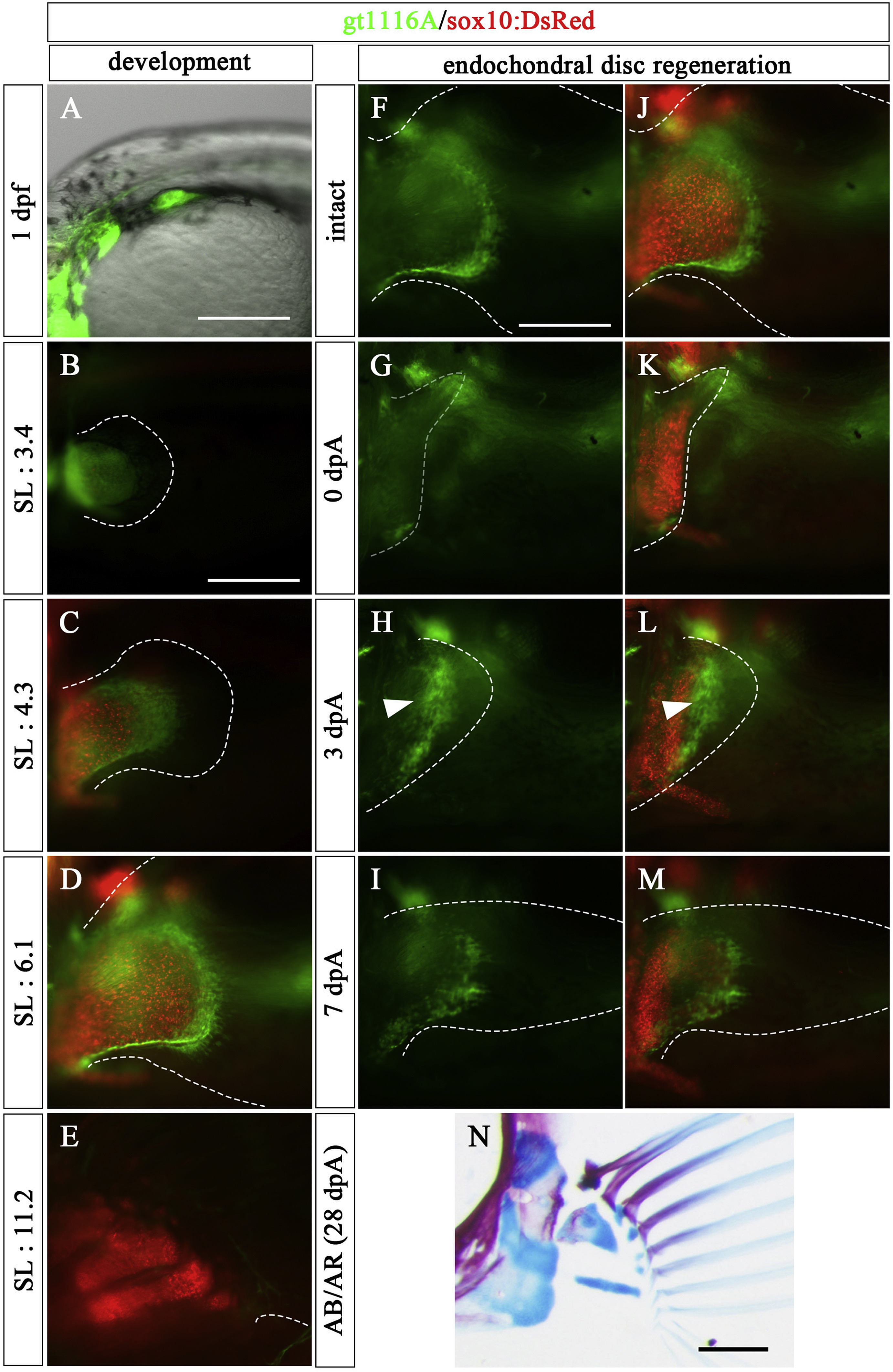
Fig. 7 Expression patterns of gt1116A in pectoral fin development and Type 1 regeneration (5.0–7.0 mm SL larvae). (A–E) Expression pattern of UAS:EGFP in the pectoral fin of a gt1116A transgenic fish observed at embryonic (1 dpf) and post-hatching stages (SL: 3.4, 4.3, 6.1, and 11.2 mm) with observation of chondrocytes using sox10:DsRed. (F–N) The amputated pectoral fin at the middle of endochondral disc in a gt1116A transgenic fish. (F–I) Expression pattern of UAS:EGFP of gt1116A. (J–M) Merged images of expression pattern of the UAS:EGFP and sox10:DsRed. White arrowheads show re-expression of UAS:EGFP at 3 dpA. (N) the pectoral fin skeletons stained with AR and AB at 28 dpA. Dashed lines indicate the outline of fins and the UAS:EGFP-positive mesenchymal cells in the regenerated region, respectively. Scale bars: 200 μm in A, B, F, and N.
Reprinted from Developmental Biology, 463(2), Yoshida, K., Kawakami, K., Abe, G., Tamura, K., Zebrafish can regenerate endoskeleton in larval pectoral fin but the regenerative ability declines, 110-123, Copyright (2020) with permission from Elsevier. Full text @ Dev. Biol.