
Fig. 8
- ID
- ZDB-FIG-200714-20
- Publication
- Sidhwani et al., 2020 - Cardiac function modulates endocardial cell dynamics to shape the cardiac outflow tract
- Other Figures
- All Figure Page
- Back to All Figure Page
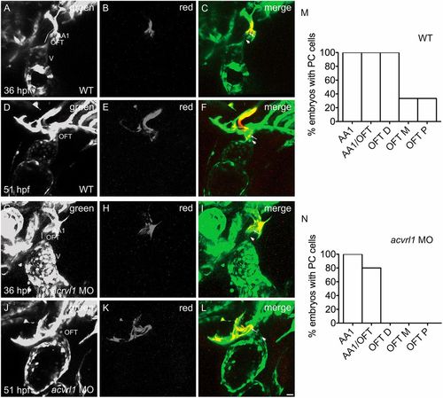
Acvrl1 promotes OFT growth by regulating endothelial addition. (A-N) Three-dimensional reconstructions depict the photoconversion of a portion of AA1 endothelium at 36 hpf (as in Fig. 2Q-S) in wild-type (WT) embryos (A-C) or acvrl1 morphants (G-I). By 51 hpf, labeled cells are routinely detected (as in Fig. 2T-V) in the WT OFT (D-F), as quantified in M (n=3; N=2). However, in acvrl1 morphants, labeled AA1 endothelial cells fail to accrete to the OFT by 51 hpf (J-L) (n=5; N=2). See also Movie 8. Arrowheads indicate examples of cells that were not labeled by photoconversion (green only) and arrows indicate examples of cells that were labeled by photoconversion (red and green). (M,N) Bar graphs (as in Fig. S1) indicate the percentage of embryos in which photoconversion of AA1 endothelium at 36 hpf resulted in detection of photoconverted (PC) cells in AA1, at the junction of AA1 and the OFT (AA1/OFT), the distal OFT (OFT D), the middle OFT (OFT M) or the proximal OFT (OFT P) at 51 hpf. Scale bar: 20 μm. |