FIGURE

Figure 6

ID
ZDB-FIG-200627-7
Publication
Weaver et al., 2020 - Hyaloid vasculature and mmp2 activity play a role during optic fissure fusion in zebrafish
Other Figures
All Figure Page
Back to All Figure Page
Figure 6

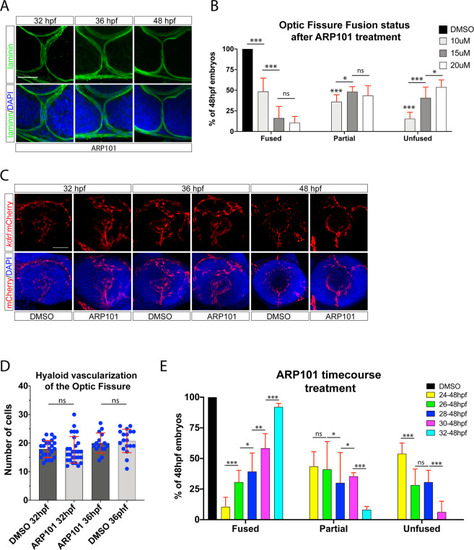
Proper timing of mmp2 activity is required for optic fissure fusion. (A) Whole mount Immunohistochemistry was used to visualize laminin (green) and DAPI (blue) in ARP101 treated Tg[rx3:GFP] embryos at 32, 36 and 48 hpf. Stacks of central-proximal regions of the OF are displayed. Scale bar = 50 μm. (B) Quantification of 48 hpf ARP101 treated embryos for fissure fusion (absence of laminin signal), partial fusion (partial retention of laminin) or failure to fuse (retention of laminin throughout the fissure). n = 41 (10 μM), 38 (15 μM), 42 (20 μM) ANOVA p < 0.0001. (C) 3D reconstructions of whole mount Tg[kdrl:mCherry] (red) embryos treated with 20 μM ARP101 from 24-32, 36 or 38 hpf. DNA was stained with DAPI (blue). Scale bar = 50 μm. (D) Quantification of the number of mCherry positive cells from 3D confocal stacks within the region of the OF after DMSO or 20 μM ARP101 treatment. Individual embryo results are depicted. (E) Quantification of 48 hpf ARP101 treated embryos for fissure fusion, partial fusion or failure to fuse after various treatment initiation times from 24-32 hpf. n = 42 (24-48 hpf), 29 (26-48 hpf), 29 (28-48 hpf), 23 (30-48 hpf), 37 (32-48 hpf). ANOVA p < 0.0001.

Expression Data

Expression Detail
Antibody Labeling
Phenotype Data
Fish:
Condition:
Observed In:
Stage: Long-pec

Phenotype Detail
Acknowledgments
This image is the copyrighted work of the attributed author or publisher, and ZFIN has permission only to display this image to its users. Additional permissions should be obtained from the applicable author or publisher of the image. Full text @ Sci. Rep.