
Fig. 2
- ID
- ZDB-FIG-200626-1
- Publication
- Sagarin et al., 2019 - Anterior Trunk Muscle Shows Mix of Axial and Appendicular Developmental Patterns
- Other Figures
- All Figure Page
- Back to All Figure Page
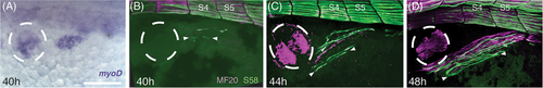
PHM differentiation begins with slow fibers elongating distant from the somites and then growing anteriorly and posteriorly. Anterior is to the left. Embryos are in dorsolateral view. Dotted circle represents the position of the pectoral fin bud. A, myoD expression at 40 hours showing the relative location of the earliest PHM fibers. B‐D, Antibody labeling for S58 (slow myosin, green) and MF20 (all muscle myosin, magenta), arrowheads indicate the ends of the most recently formed muscle fibers at each stage, somite 4 and somite 5 are indicated (S4, S5). B, 40 hours Myosin at somites 2‐6 shows the first PHM fibers separate from the axial myotome. C, 44 hours Myosin at somites 1‐6 showing the PHM expanded anteriorly and posteriorly, and the second segment of the PHM forming at ventral somite 6. D, 48 hours Myosin at somites 1‐6 (S4 and S5 labeled) showing fiber striations and further expansion. Scale bar in A is 100 μm. Scale bar in D, for B to D, is 50 μm |