
Fig. s9
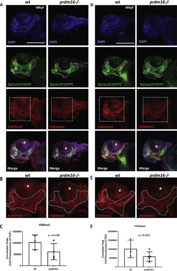
Loss of prdm16 reduces trimethylation of histone H3 substrates in zebrafish pharyngeal arch derived craniofacial structures. (A-F) Wildtype or prdm16−/− zebrafish embryos crossed into the Tg(sox10:EGFP) transgenic line were collected at 48 hpf and whole mount immunofluorescence was performed for trimethylated H3 lysine 9 (A-C) or trimethylated H3 lysine 4 (D-F). Shown are ventral confocal max projection images of the head. (B, E) Zoomed images of H3K9me3 or H3K4me3. (C, F) Quantification of the corrected total mean fluorescence intensity of H3K9me3 (C) or H3K4me3 (F) in the pharyngeal arch-derived craniofacial structures outlined with dotted line in (B) and (E) from 3 to 5 different embryos for each genotype and each mark. Abbreviation: eye (e). Scale bar 150 μm p values shown above, Student’s t-test. |
Reprinted from Developmental Biology, 461(2), Shull, L.C., Sen, R., Menzel, J., Goyama, S., Kurokawa, M., Artinger, K.B., The conserved and divergent roles of Prdm3 and Prdm16 in zebrafish and mouse craniofacial development, 132-144, Copyright (2020) with permission from Elsevier. Full text @ Dev. Biol.