|
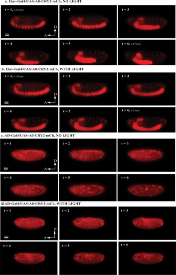
Light-induced Aβ aggregation in <italic>Drosophila</italic> embryos resulted in distinct developmental defects.(a) Still images from a 24 hr recording of Elav-Gal4/UAS-Aβ- CRY2-mCh aggregates in a Drosophila embryo without blue light illumination resulted in normal development. Time stages t = 1–6 showed embryonic development stages 12–13 (~440–620 min after fertilization), where germ band retraction starts and ends respectively. Germ band was 5.125 mm at t = 1, and retracted to 2.875 mm by t = 6. (b) Still images from a 24 hr recording of Elav-Gal4/UAS-Aβ-CRY2-mCh aggregates in a Drosophila embryo with blue light illumination every 2.5 min resulted in the arrest of embryogenesis during late germ band retraction stages. Time stages t = 1–6 showed embryonic development from late stage 10 to early stage 12 (~300–460 min after fertilization), where germ band retraction begins (stage 12). Germ band was 5.125 mm at t = 1, and retracted only to 4.25 mm by t = 6. (c) Still images from a 24 hr recording of uniform expression AD-Gal4/UAS-Aβ-CRY2-mCh aggregates in a Drosophila embryo without blue light illumination resulted in normal development. Time stages t = 1–6 showed embryonic development stages 14–16 (~620–900 min after fertilization), where dorsal closure of midgut and epidermis and shortening of ventral nerve cord occurs. In this case, the ventral nerve cord shortening was not clearly visible. (d) Still images from a 24 hr recording of AD-Gal4/UAS-Aβ-CRY2-mCh aggregates in a Drosophila embryo with blue light illumination at every 2.5 min resulted in the arrest of embryogenesis at dorsal closure stages. Time stages t = 1–6 corresponded to embryonic development stage 14 (approximately 620–680 min after fertilization), where dorsal closure begins.
|