FIGURE

Figure 1

ID
ZDB-FIG-200421-77
Publication
Sensi et al., 2020 - Recellularized Colorectal Cancer Patient-derived Scaffolds as in vitro Pre-clinical 3D Model for Drug Screening
Other Figures
All Figure Page
Back to All Figure Page
Figure 1

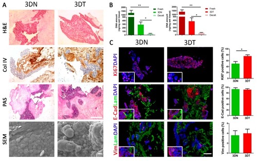
Characterization of matched 3DN and 3DT HT29-recellularized samples. (A) histological characterization of sections stained with hematoxylin and eosin (H&E), collagen IV staining (Col IV), and periodic acid-Shiff (PAS) (scale bar = 200 µm). Scanning electron microscopy (SEM) analysis performed on 3DN and 3DT (Scale bar = 10 µm). (B) DNA amount quantification in fresh samples, after decellularization and 5 days of culture after seeding of HT29 cells, in both 3DN and 3DT. (C) IF staining in 3DT and 3DN and relative quantifications: Ki67, as proliferation marker; E-cadherin, as epithelial marker; vimentin, as mesenchymal marker; laminin to highlight basement membrane; DAPI to counterstain the nuclei (scale bar = 100 µm) (* p-value < 0.05; ** p-value < 0.01).

Expression Data

Expression Detail
Antibody Labeling
Phenotype Data

Phenotype Detail
Acknowledgments
This image is the copyrighted work of the attributed author or publisher, and ZFIN has permission only to display this image to its users. Additional permissions should be obtained from the applicable author or publisher of the image. Full text @ Cancers