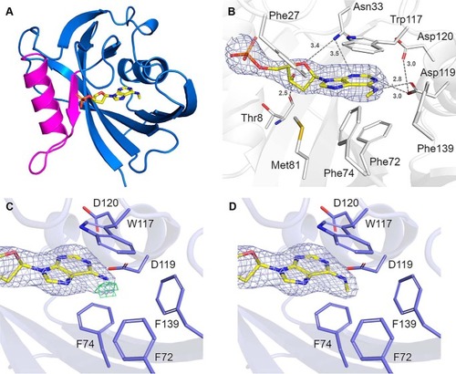
Crystal structure of hMTH1 in complex with N6-methyl-dAMP.A, overall structure of hMTH1 in ribbon representation, colored blue. The Nudix motif is colored magenta. N6-methyl-dAMP is presented as a stick model. B, the active site hydrogen bond network of hMTH1 with the reaction hydrolysis product N6-methyl-AMP (N6-met-AMP), with the 2Fo − Fc composite omit map contoured at 1.0 σ. Important binding residues and residues of the hydrophobic pocket are depicted as sticks; C atoms are colored white, O atoms red, N atoms blue, and S atoms gold. N6-methyl-dAMP is presented as a stick model; C atoms are colored yellow, O atoms red, N atoms blue, and P atoms orange. Hydrogen bond interactions are shown as dashed lines with bond distances indicated in Angstroms (Å). C and D, refinement of hMTH1 structure. The ligands (C) dAMP and (D) N6-methyl-dAMP were modeled into hMTH1 in Coot (58) following which the structures were refined using Refmac5 (59). The 2Fo − Fc electron density maps around the ligands following refinement are contoured at 1.0 σ (blue) and the Fc − Fc electron density maps are contoured at −2.5 σ (red) and +2.5 σ (green). Figures were produced with PyMOL (version 2.1.1, Schrödinger). Single letter amino acids are used in the figure.
|
