
Fig. 2
- ID
- ZDB-FIG-200406-283
- Publication
- Ohata et al., 2020 - An Activity-Based Methionine Bioconjugation Approach To Developing Proximity-Activated Imaging Reporters
- Other Figures
- All Figure Page
- Back to All Figure Page
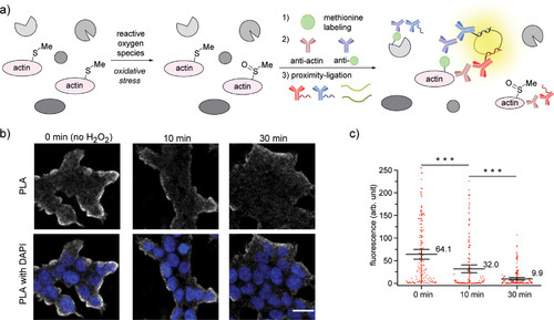
“Turn-off” detection of oxidative stress in cells via methionine proximity-activated imaging reporter (Met-PAIR) on β-actin. (a) Schematic for Met-PAIR on β-actin. (b) Confocal images of β-actin Met-PAIR on HEK293T cells preincubated with H2O2 (1 mM) for 0, 10, or 30 min, washed, and labeled with alkyne-tagged oxaziridine (Ox4, 20 μM) for 20 min. Ox4-labeled β-actin was further functionalized with Oregon Green-azide, and proximity-ligation assay (PLA) stain was performed with mouse anti-β-actin antibody and rabbit anti-Oregon Green antibody. Gray: PLA staining. Blue: DAPI nuclear staining. Scale bar: 20 μm. (c) Whisker plots for the confocal images in (b). Each dot represents fluorescence intensity of single cells. Whisker and center line represent 95% confidence interval and mean intensity, respectively. The mean value is shown near the center line. The quantification was conducted by imaging 4 regions in each of 2 independent biological replicates (total 8 cell images). The number of the quantified cells: 119 (0 min), 131 (10 min), and 157 (30 min). ***P < 0.001, Student’s t test. |