
Fig. 3
- ID
- ZDB-FIG-200331-4
- Publication
- Bensimon-Brito et al., 2019 - TGF-β Signaling Promotes Tissue Formation during Cardiac Valve Regeneration in Adult Zebrafish
- Other Figures
- All Figure Page
- Back to All Figure Page
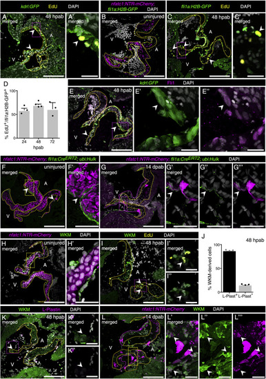
New Valve Cells Differentiate from Endothelial Cells and Kidney Marrow-Derived Cells (A and A′) EdU detection and kdrl:GFP expression in 48 hpab valves (arrowheads point to double-positive cells; n = 4). (B) Uninjured valve showing absence of fli1a:H2B-GFP expression in VICs (n = 4). (C and D) Characterization (C and C′; n = 4) and quantification of the percentage (D) of EdU+ cells positive for fli1a:H2B-GFP expression. Boxed areas shown in (A′) and (C′). (E) Co-localization of kdrl reporter with Fli1 antibody (arrowheads; n = 4). Boxed areas shown in E′ and (E″). (F–G″′) Cre/lox lineage tracing experiments show a few endothelial-derived nfatc1+ cells (arrowheads) in uninjured valves (F and F′; n = 6) and contribution of endothelial cells to nfatc1+ VICs (arrowheads) at 14 dpab (G–G″′; n = 9). Boxed areas shown in (F′) and (G′–G″′). Yellow and pink dashed lines delineate the old and new valve leaflets, respectively. (H–I″) WKM-derived cells in uninjured valves (H and H′; n = 3) and positive for EdU (I–I″) at 48 hpab. (J–K″) Quantification (J) and localization (K–K″) of WKM-derived cells negative for the immune cell marker L-Plastin. Boxed areas shown in (H′), (I′), (I″), (K′), and (K″). (L–L″′) Contribution of circulating WKM-derived cells to new nfatc1+ cells (arrowheads) at 14 dpab (n = 4). Boxed areas shown in (L′–L″′). A, atrium; V, ventricle. Scale bars: (A), (B), (C), (E), (F), (G), (H), (I), (K), and (L) 100 μm; (A′), (C′), (E′ and E″), (F′), (G′–G″′), (H′), (I′–I″), (K′ and K″), and (L′–L″′) 20 μm. |
Reprinted from Developmental Cell, 52(1), Bensimon-Brito, A., Ramkumar, S., Boezio, G.L.M., Guenther, S., Kuenne, C., Helker, C.S.M., Sánchez-Iranzo, H., Iloska, D., Piesker, J., Pullamsetti, S., Mercader, N., Beis, D., Stainier, D.Y.R., TGF-β Signaling Promotes Tissue Formation during Cardiac Valve Regeneration in Adult Zebrafish, 9-20.e7, Copyright (2019) with permission from Elsevier. Full text @ Dev. Cell