
Fig. 3
- ID
- ZDB-FIG-200326-46
- Publication
- Jurynec et al., 2019 - The Paf1 Complex and P-TEFb have reciprocal and antagonist roles in maintaining multipotent neural crest progenitors
- Other Figures
- All Figure Page
- Back to All Figure Page
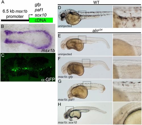
Paf1 is required cell-autonomously in the NC lineage for melanophore development. (A) Schematic representation of a msx1b promoter construct used to express gfp, paf1 or sox10 cDNAs. (B) Endogenous msx1b expression in an 11.5 hpf embryo detected by whole-mount in situ hybridization. (C) GFP (detected by immunohistochemistry) is expressed mosaically, but only within the normal msx1b expression domain of an 11.5 hpf embryo injected at the one-cell stage with 50 pg msx1b::GFP plasmid DNA. (D-H) 48 hpf control and DNA-injected embryos. (D) Wild-type embryo exhibiting normal distribution and morphology of melanophores. Uninjected alnz24 mutant embryos (E) and alnz24 mutants injected with msx1b::GFP plasmid DNA (F) completely lack NC-derived melanophores. (G) alnz24 mutants injected with msx1b::paf1 plasmid DNA have widely distributed melanophores with normal stellate morphology. (H) Expression of sox10 in the msx1b expression domain fails to rescue melanophore development; abnormal pigment cells were occasionally found in the heads of plasmid-injected mutant embryos. (B,C) Dorsal views of flat-mounted embryos, with anterior towards the left. D-H are lateral views of entire embryos (anterior towards the left); boxed regions are shown at higher magnification to the right of each whole-embryo view. |