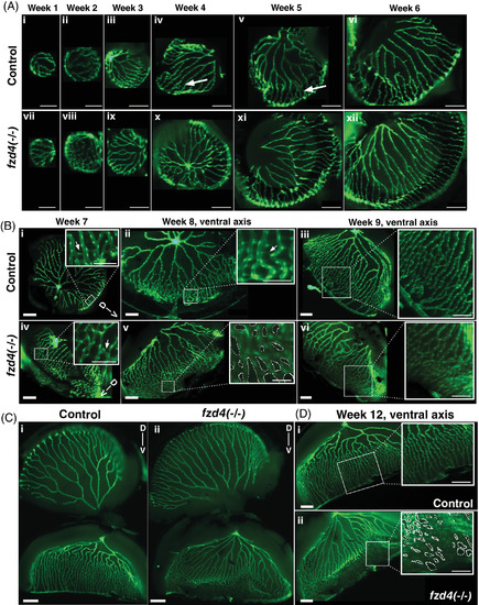
Developmental progression of zebrafish retinal vasculature in control and fzd4 fish. A, Weeks 1‐6. The retinal vasculature engulfs the entire lens as early as week 1. A few anastomoses are detected as early as week 4 in both control (arrows in iv, v) and fzd4 mutants (not shown). B, Weeks 7‐9. Ventral asymmetry can now be detected in both control and fzd4 mutants, in which the vessels on the ventral side are more closely opposed compared to those on the dorsal side. At week 8, ventral fusion is more pronounced in fzd4 mutants than in controls. Arrows in B‐i and B‐iv insets indicate sprouts. C‐D, Week 12. In, C, control and mutant retinas are shown whole to show the difference in dorsal to ventral arrangement of their vessels. In, D, only the ventral side from, C, is shown to emphasize the difference in vessel morphology and fusion between control and mutant eyes (inset shows severe fusion in a lateral section of the eye). Scale bars in, A, i‐ii, and vii‐viii is 100 μm, and A, ix‐xii is 200 μm. Scale bars in, B, i‐vi is 200 μm and in insets 100 μm. Scale bar in, C, is 500 μm. Scale bars in, D, are 200 μm, inset are 100 μm. Dotted circles in inset in, B‐v and D‐ii, highlight the spaces between blood vessels to emphasize the degree of abnormal anastomoses
|
