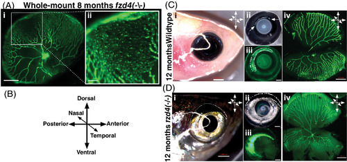
Asymmetric retinal vasculature distribution and ventral restriction of the fzd4 vascular phenotype. A, Whole mount retina from an 8‐month old fzd4 mutant. GFP marks endothelial cells (fli:eGFP). Note abnormally high vessel density and apparent fusion of nearby vessels to form disorganized endothelial sheets. Scale bar 500 μm and inset 100 μm. B, Orientation of the eyes with respect to the body axes of the fish in C, D. C‐D, Orientation of a right eye from control (C) and fzd4 (D) zebrafish using external morphology before enucleation (i), after enucleation with brightfield in C‐ii, (arrowheads depicts a bright band on the dorsal side above the lens in casper fish and in D‐ii, (dotted circle highlights a dark dorsal band in fzd4 fish, and with fluorescence in C‐iii and D‐iii (dotted circles depict a bright fluorescent band on the dorsal posterior surface in C‐iii, and D‐iii), and during dissection of the tissue (C‐iv and D‐iv). Scale bar in Ci and Di is 1 mm and in Cii‐iv, Dii‐iv is 500 μm
|

