
Fig. 2
- ID
- ZDB-FIG-200319-14
- Publication
- Usui et al., 2019 - The minimal gap-junction network among melanophores and xanthophores required for stripe-pattern formation in zebrafish
- Other Figures
- All Figure Page
- Back to All Figure Page
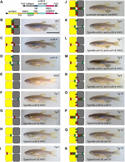
Mutant and transgenic fish lines, and reconstructed gap junction networks. (A) Plasmid construct designs: mitfa promoter (A; upper line) and aox5 promoter (A; lower line) were used for pigment-cell-specific gene expression. tTA/TRE was used to enhance aox5 promoter activity, and an ubipro (ubiquitinb promoter)-EGFP cassette was used to simplify the genotyping of fish embryos. The fragments were cloned into a pTol2 plasmid, and each plasmid was used to generate transgenic zebrafish. (B-R) The reconstructed gap junction network (left) and a representative photograph of a fish from the corresponding line (right). Wild-type (B; WT), cx39.4−/− (C; luchs), cx41.8−/− (D; leopard) and double-knockout mutant (E; WKO), and transgenic zebrafish lines in WKO background (F-N; Tg-1 to Tg-9), cx39.4−/− background (O,P; Tg-10 and Tg-11) and cx41.8−/− background (Q,R; Tg-12 and Tg-13). mitfa promoter (F,G,O,Q) and aox5 promoter (H,I,P,R) were used to induce cx39.4 or cx41.8 in pigment cells. Double (K-N) and quadruple (J) transgenic lines were generated by means of crossing among mutant and transgenic lines. Gene expression in unexpected cells was monitored using IRES-H2BRFP fluorescent protein (Fig. S2). Scale bar: 10 mm. |